
Interests
Research interests · Vascular mechanobiology
What makes a blood vessel
vulnerable to failure?
I investigate how vascular cells maintain the matrix that enables a vessel to withstand load, and how disruption of this system creates mechanical vulnerability. The aim is to connect cellular regulation, spatial matrix architecture, and tissue mechanics in models that explain and eventually predict failure.
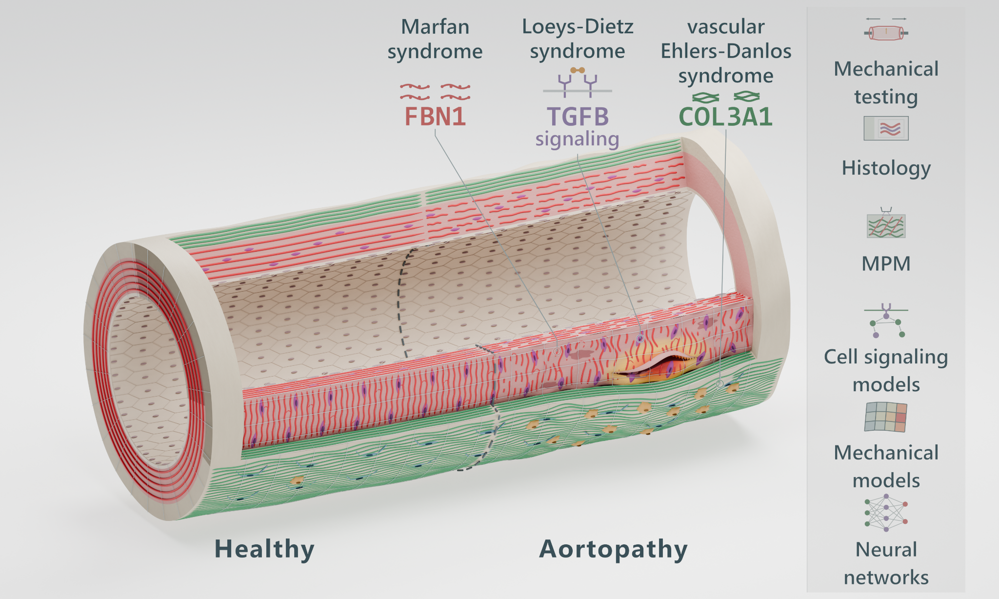
Which cellular programs regulate matrix integrity?
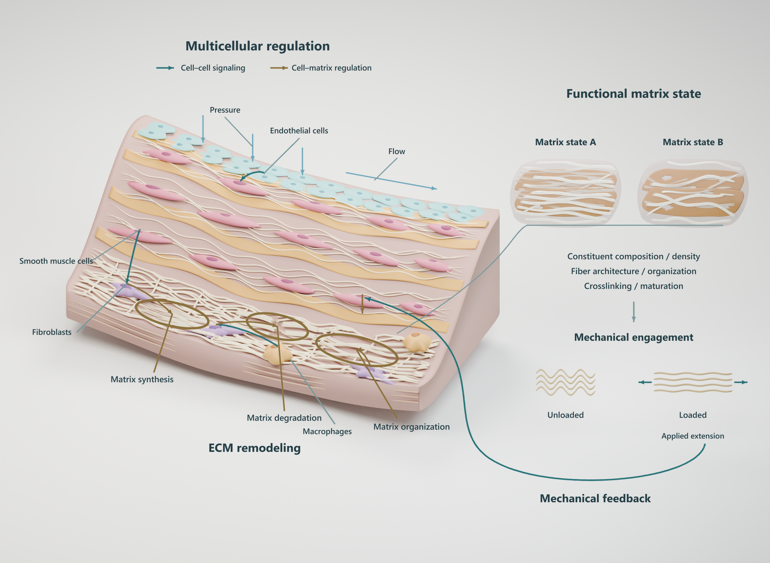
Smooth muscle cells, fibroblasts, endothelial cells, and immune cells collectively regulate matrix production, degradation, and maintenance. Genetic disease, altered signaling, and pressure loading provide complementary ways to investigate disruption of this control system.
The question: Which changes in cell state produce a mechanically consequential matrix phenotype?
The approach: Connect spatial molecular profiling and targeted protein measurements with the matrix actually present in the tissue.
Cell signalingSpatial profilingCell–matrix interactionsWhich matrix changes create mechanical vulnerability?
Collagen or elastin abundance alone cannot describe how a vessel bears load. Matrix density, fiber organization, recruitment, crosslinking, and lamellar integrity determine which constituents are available and engaged as the wall deforms.
The question: When does remodeling preserve load sharing, and when does it concentrate demand in vulnerable regions?
The approach: Spatial microstructural characterization and multiphoton imaging under controlled loading, paired with biochemical and histologic measurements.
Functional matrix densityCollagen recruitmentSpatial microstructureHow does matrix state determine tissue strength?
Mechanical testing provides the bridge between a measured matrix phenotype and the tissue's ability to resist failure. My work combines biaxial mechanics, inflation-based testing, and microstructural imaging to connect constituent engagement with deformation and strength.
The question: Which local structural features explain failure initiation and propagation beyond geometry or bulk composition?
The approach: Test fresh vascular tissue under controlled loading and compare deformation and failure with spatially matched structural measurements.
Biaxial mechanicsInflation testingFailure initiationStructure becomes function.
Cell-driven changes in matrix state alter constituent engagement and load sharing. The resulting mechanical demand can help distinguish adaptation from vulnerability.
Prior work links proteolytic changes in aortic tissue density with stiffness and tensile strength.
Read the publications →Can coupled models predict adaptation and failure?
My long-term aim is to couple cell-signaling network models with constrained-mixture models of vascular growth and remodeling. Predicted changes in matrix turnover and organization would update constituent properties, tissue mechanics, and vulnerability over time.
The question: Given a specific perturbation, can we predict how the tissue's mechanical state evolves and whether it moves toward adaptation or failure?
The approach: Graph-based neural networks and mechanistically constrained surrogate models, tested against independent experimental measurements.
Cell-signaling networksConstrained-mixture modelingAI-enabled predictionMechanistic connections
Perturbations and tissue response
Marfan · FBN1
Fibrillin dysfunction
Altered microfibrillar structure and cell–matrix signaling provide the starting context.
Realized remodeling
Measure lamellar integrity, collagen organization, and the matrix available to bear load.
Constituent engagement
Test how altered architecture changes recruitment and the distribution of mechanical demand.
Failure susceptibility
Determine which measured changes explain strength and the onset of tissue failure.
TGF-β signaling
SMC signaling disruption
Altered TGF-β receptor signaling offers a way to study changes in smooth muscle cell regulation.
Production and turnover
Connect molecular programs with measured matrix abundance, organization, and maintenance.
Changing load sharing
Evaluate how the resulting matrix state shifts constituent engagement during deformation.
Adaptation or failure
Test whether a coupled signaling and remodeling model explains progression toward mechanical vulnerability.
vEDS · COL3A1
Type III collagen dysfunction
COL3A1-related disease provides a context for investigating impaired matrix integrity.
Effective load-bearing matrix
Characterize collagen organization and the matrix that remains mechanically functional.
Local demand and strength
Assess whether spatial matrix changes concentrate demand or reduce resistance to failure.
Mechanistic prediction
Evaluate which structural and mechanical measures improve experimentally validated failure predictions.
Long-term objective
Predict mechanical state and failure vulnerability.
The goal is an experimentally grounded explanation of how cellular dysfunction changes vascular integrity over time, providing a mechanistic foundation for future risk assessment.
-
I hope to contribute to understanding the mechanical and microstructural components that lead to vascular disease. I primarily study abdominal aortic aneurysms but also cerebral aneurysms, thoracic aortic aneurysms, and deep vein thrombosis.
I work to improve methods of diagnosis by utilizing patient specific metrics rather than a one-size-fits-all maximum diameter criterion. A patient-specific holistic approach of multiple variables yields higher chances of properly assessing risk of aneurysms.
-
I am passionate about designing new experimental techniques to analyze vascular tissue mechanically. Utilizing techniques like stereo digital image correlation, uniaxial extension, biaxial extension, and fluid-pressurization are just a few of the techniques I utilize in my research.
-
I enjoy coding (primarily MATLAB and Python) software that automates and improves medical image analysis and experimental techniques.
I also like to design software that finds exciting new ways to show and present data. Including augmented and artificial reality, which is becoming more and more prevalent in the medical field.
-
I discovered my love of teaching in the fall of 2021 when I served as a teaching assistant for Biomechanics 2 for Dr. Richard Debski. Since then, I also served as a teaching assistant for Biomechanics 1 in spring of 2022 for Dr. Spandan Maiti.
These 2 professors, Dr. Debski and Dr. Maiti, have both inspired my love of teaching and my teaching style, which is highly encouraging of students to apply their knowledge through projects and labs.
